


Microplates

Choosing the right microplate is a critical, often overlooked, part of an assay. The right microplate helps provides valuable data, whereas the wrong microplate can lead to missed or inaccurate data leading to missed project timelines and ultimately higher costs.
New technologies for drug development are rapidly advancing. These technologies have given rise to a variety of applications that require their own unique consumables. At Revvity, we provide microplates that help you discover better. Covering a vast array of application areas, Revvity microplates are available in different well formats, plastic types, coatings, and colors – clear, white, black, gray. Our microplates have been engineered to deliver the highest quality data for applications ranging from high throughput screening to cellular imaging. Discover the difference a better microplate makes:
- Footprint dimensions meet the SBS industry standard, guaranteeing compatibility with microplate-based instrumentation
- Carefully monitored microplate planarity to ensure superb data and image acquisition
- Custom microplate services including barcoding, custom packaging, as well as plate surface coatings to fit your application
- Full range of validated reagents and instruments to complement microplates
For research use only. Not for use in diagnostic procedures.
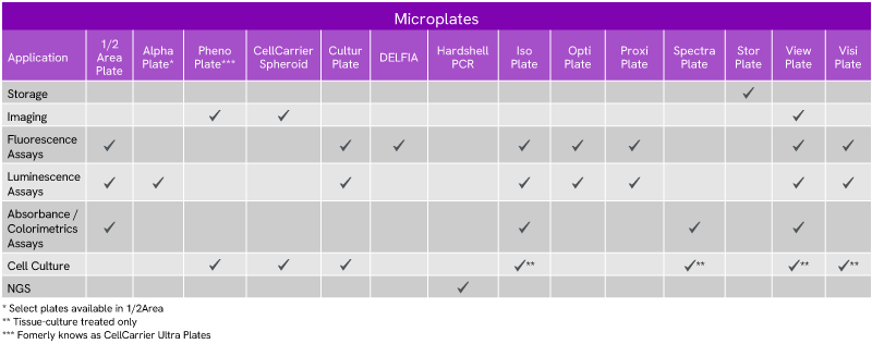

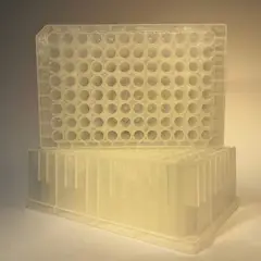
Storage
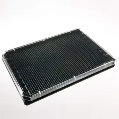
Microplates for storing reagents, biomolecules, or other samples.
Microplates for storing reagents, biomolecules, or other samples.

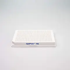
Cell imaging
Microplates for microscopy and high-content screening offering the lowest plate bottom for superior image acquisition.
Microplates for microscopy and high-content screening offering the lowest plate bottom for superior image acquisition.

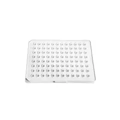
Assays
Microplates for TR-FRET, HTRF, Alpha, Radiometric, luminescence, fluorescence, and others for general research.
Microplates for TR-FRET, HTRF, Alpha, Radiometric, luminescence, fluorescence, and others for general research.

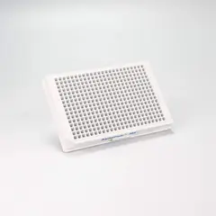
Cell culture
Microplates designed to improve consistency and reliability of cell growth for general cell culture as well as 3D cell culture.
Microplates designed to improve consistency and reliability of cell growth for general cell culture as well as 3D cell culture.

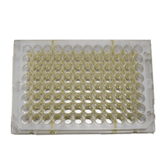
Genomic analysis
Microplates for genomic analysis used in sample preparation workflows.
Microplates for genomic analysis used in sample preparation workflows.

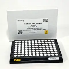
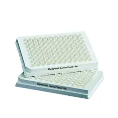
Radiometric
Microplates for a range of high-throughput radiometric assays.
Microplates for a range of high-throughput radiometric assays.


Filters
View Product Listing Solr Page, display Block: Product Type
View Product Listing Solr Page, display Block: Application
View Product Listing Solr Page, display Block: Brand
View Product Listing Solr Page, display Block: Coating Treatment
View Product Listing Solr Page, display Block: Color
View Product Listing Solr Page, display Block: Detection Method
View Product Listing Solr Page, display Block: Product Group
View Product Listing Solr Page, display Block: Radioisotope
View Product Listing Solr Page, display Block: Sample Type
View Product Listing Solr Page, display Block: Shape
View Product Listing Solr Page, display Block: Sterility
View Product Listing Solr Page, display Block: Technology Type
View Product Listing Solr Page, display Block: Type
View Product Listing Solr Page, display Block: Volume
View Product Listing Solr Page, display Block: Well Format
View Product Listing Solr Page, display Block: Well Shape
1 - 25 of 170 Products and Services
.jpg?format=webp&width=240)


Filters
View Resource Library Media Listing, display Block: Resource Type