Strict R Inducible CRISPRaレンチウイルスシステム
Dharmacon™ Strict R™ Inducible CRISPRaレンチウイルスシステムは、精密かつ調整可能な遺伝子活性化を可能にします。本システムは、標的遺伝子の発現を高い特異性と効率で制御的に上昇させることができる、初のプラットフォームです。Tet On 3GシステムとFKBP12由来デグロンシステムの組み合わせにより、低分子化合物に応答したdCas9 VPRの誘導的活性化および分解が可能となり、不要なバックグラウンド発現を最小限に抑えます。
精密な遺伝子活性化を可能にする二重誘導制御:仕組み
System OFF:ドキシサイクリンおよびShield1が存在しない場合、本システムは不活性状態を維持します。TRE3Gプロモーターからの残存的な転写によりデグロンタグ付きdCas9 VPRが産生された場合でも、当該タンパク質は速やかにプロテアソーム分解を受けるため、意図しないバックグラウンド活性化は最小限に抑えられます。
System ON:ドキシサイクリンの添加によりTet On 3Gトランスアクチベーターが活性化され、dCas9 VPRの強力な発現が誘導されます。さらにShield1を添加することで、発現したdCas9 VPRタンパク質が安定化され、蓄積が可能となり、精密な遺伝子活性化が実現されます。
Dharmacon Strict R Inducible CRISPRaレンチウイルスベクターの模式図

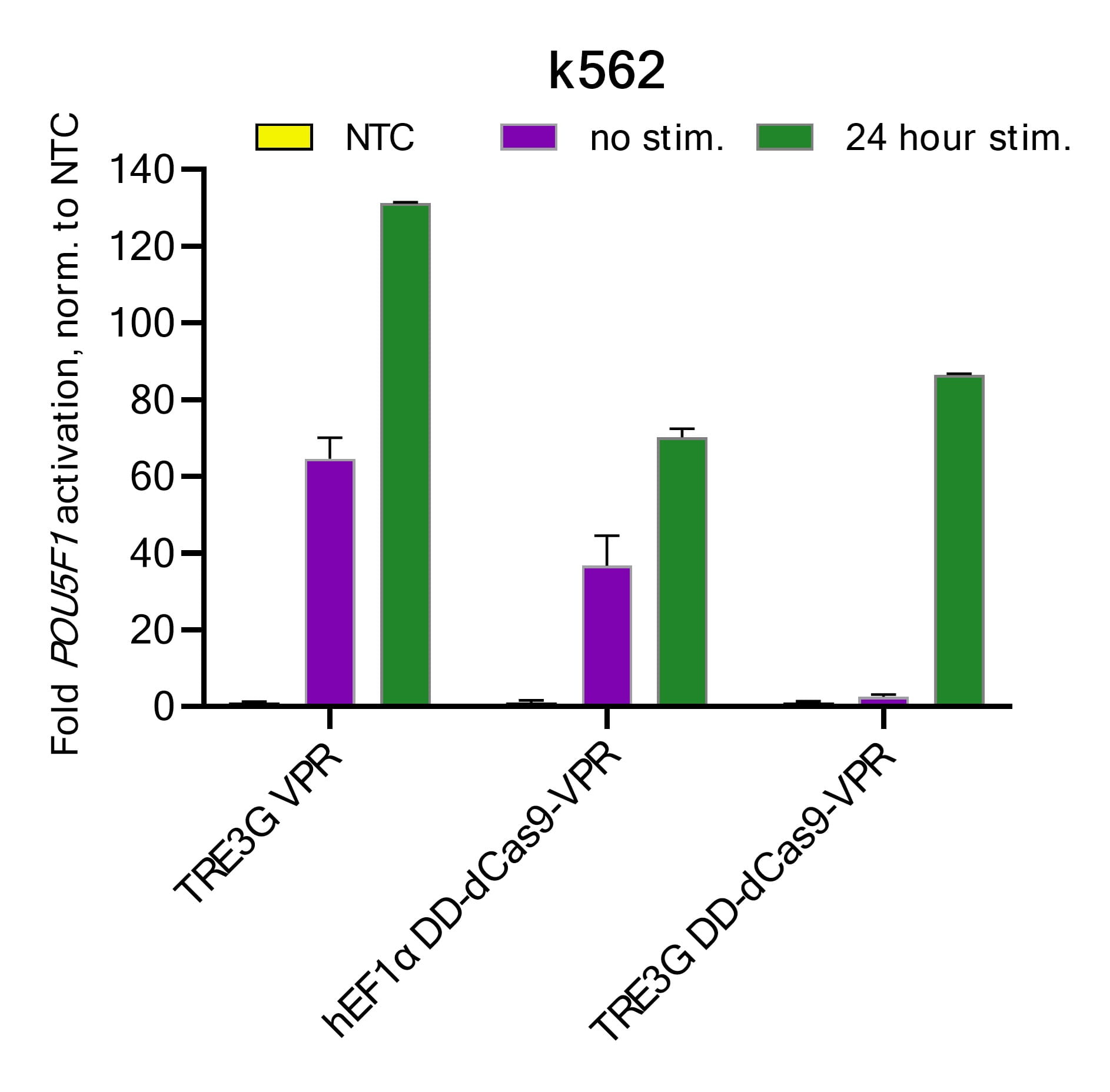
Dharmacon Strict R 誘導型 CRISPRaレンチウイルスシステムによる転写レベルおよび翻訳後レベルでの制御

Strict R Inducible Lentiviral CRISPRaシステムの模式図を示しています。ドキシサイクリンおよびShield1非存在下では、本システムは「OFF」状態となります。TRE3Gプロモーターからのリーキーな転写により、FKBP12由来の不安定化ドメイン(デグロン)と融合したdCas9 VPRが翻訳されますが、当該タンパク質は速やかにプロテアソーム分解を受けます。ドキシサイクリンの添加によりTRE3Gプロモーターからの強力な転写が誘導され、さらにShield1の添加によってdCas9 VPRが安定化されることで、遺伝子特異的sgRNA存在下において高効率な標的遺伝子活性化が可能になります。本図はBioRender.comを用いて作成されています。
Highlights
- 低分子誘導による可逆的な標的遺伝子活性化
- dCas9‑VPR融合タンパク質による高効率な転写活性化と低いオフターゲット影響
- ドキシサイクリンおよびShield1添加により迅速なdCas9‑VPR発現誘導を可能にするTet‑デグロンシステム
- 単一のレンチウイルスベクターによる発現で、既存の実験ワークフローに容易に統合可能
- 遺伝子発現解析、機能ゲノミクス、スクリーニング、治療応用研究など幅広い用途に対応
- 高品質・精製済みレンチウイルス粒子を低細胞毒性で直接導入可能(力価:≥1 × 10⁷ TU/mL)
- ブラスチジン選択またはEGFP蛍光レポーターの選択肢を用意