- Products
- 遺伝子発現調節試薬
- RNA干渉
Accell eGFP Control siRNA
siRNA delivery without a transfection reagent

Accell eGFP Control siRNA is designed to target cloning vector pEGFP-N1 (Genbank accession # U55762) and has 100% homology with over 100 additional cloning vectors and transgenic eGFP sequences. Expressed green fluorescent protein (eGFP) is a reporter molecule for monitoring gene expression and protein localization in vivo and in real time. Accell eGFP control siRNA reagents silence expression of the GFP mRNA leading to a detectable decrease of fluorescence observed by fluorescent microscopy or by FACS analysis.
Highlights
- Accell siRNA enters cells without the need for transfection reagents, virus (or viral vectors), or instruments
- Novel siRNA modifications facilitate uptake, stability, specificity, and knockdown efficiency
- Target sequence: 5’ GCCACAACGTCTATATCAT 3’
Experimental considerations
- Accell siRNA works at a higher concentration than conventional siRNA; recommended 1 µM working concentration
- Delivery may be inhibited by the presence of BSA in serum. Optimization studies with serum-free media formulations (Accell Delivery Media) or < 2.5% serum in standard media is recommended
- Full-serum media can be added back after 48 hours of incubation. Optimal mRNA silencing is typically achieved by 72 hours or up to 96 hours for protein knockdown
eGFP expression silenced by Accell eGFP siRNA

FKHR-U2OS cells (BioImage) were treated with 1 µM Accell eGFP Control siRNA or Accell Non-targeting siRNA #1 in Accell delivery media. Reduction of eGFP expression is visualized using Cellomics ArrayScan VTI (green= GFP expression; blue= nuclear Hoechst dye).
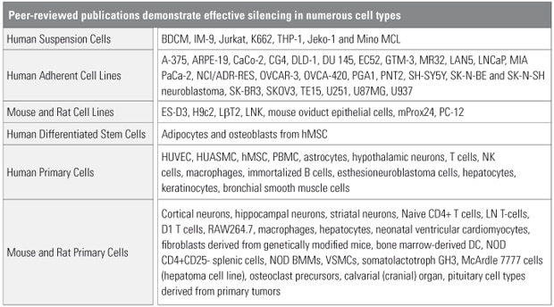
Cell types demonstrating effective silencing with Accell siRNA

Internal validation and peer-reviewed publications report numerous successes with difficult-to-transfect cell types. See the References tab for a list of publications.
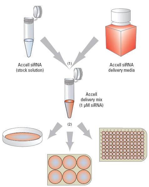
The Accell siRNA application protocol simplifies targeted gene knockdown

(1) Combine Accell siRNA with Accell delivery media (or other low- or no-serum media). (2) Add Accell delivery mix directly to cells, and incubate for 72 hours.
View the published references citing successful Accell siRNA application.
Accell siRNA reagents in neuronal cells
- A. Vagnoni et al., Calsyntenin-1 mediates axonal transport of the amyloid precursor protein and regulates Aß production. Human Molecular Genetics.21, 13 2845–2854 (2012). [rat primary cortical neurons (E18)]
- S. Suzuki et al., Differential Roles of Epac in Regulating Cell Death in Neuronal and Myocardial Cells.J. Biol. Chem.285, 24248-24259 (Jul 2010). [primary mouse cortical neurons (E15-17)]
- A. M. Dolga et al., TNF-alpha-mediates neuroprotection against glutamate-induced excitotoxicity via NF-kappaB-dependent up-regulation of K2.2 channels.J. Neurochem.107(4), 1158-1167 (November 2008). [mouse primary cortical neurons]
- U. Dreses-Werringloer et al., A Polymorphism in CALHM1 Influences Ca2+ Homeostasis, Ab Levels, and Alzheimer’s Disease Risk.Cell.133(7), 1149-1161 (27 June 2008). [SHSY-5Y; human neuroblastoma]
- J. Sebo et al., Requirement for Protein Synthesis at Developing Synapses.J. Neurosci.29(31), 9778-9793 (5 August 2009). [rat primary hippocampal neurons]
- P. Mergenthaler et al., Mitochondrial hexokinase II (HKII) and phosphoprotein enriched in astrocytes (PEA15) form a molecular switch governing cellular fate depending on the metabolic state.PNAS USA.109(5), 1518-1523 (31 January 2012). [extended duration silencing in rat primary cortical neurons]
Accell siRNA reagents in immunological cells
- D. Smirnov et al., Genetic Analysis of Radiation-induced Changes in Human Gene Expression.Nature.459(7246), 587-591 (28 May 2009). [immortalized B cells]
- V. Saini et al.,CXC Chemokine Receptor 4 Is a Cell Surface Receptor for Extracellular Ubiquitin.J. Biol. Chem.285(20), 15566-15576 (14 May 2010). [THP-1 monocytes]
- J. W. Perry et al., Endocytosis of Murine Norovirus 1 into Murine Macrophages Is Dependent on Dynamin II and Cholesterol.J. Virol.84(12), 6163-6176 (June 2010). [murine macrophages]
- M. Steenport et al., Matrix Metalloproteinase (MMP)-1 and MMP-3 Induce Macrophage MMP-9: Evidence for the Role of TNF-a and Coclooxygenase-2.J. Immunology.183(12), 8119-8127 (15 December 2009) [RAW264.7 macrophages, doi:10.4049/jimmunol.0901925]
- C. B. Lai et al., Creation of the two isoforms of rodent NKG2D was driven by a B1 retrotransposon insertion.Nucleic Acids Res. (2009). [mouse NK cell line, gkp174]
- N. Mookherjee et al., Intracellular Receptor for Human Host Sefense Peptide LL-37 in Monocytes.J. Immunol.183(4), 2688-2696 (15 August 2009). [THP-1; human monocytes]
- D. Smirnov et al., Genetic Analysis of Radiation-induced Changes in Human Gene Expression.Nature.459(7246), 587-591 (28 May 2009). [immortalized B cells]
- A. M. McElligott et al., The Novel Tubulin-Targeting Agent Pyrrolo-1,5-Benzoxazepine-15 Induces Apoptosis in Poor Prognostic Subgroups of Chronic Lymphocytic Leukemia.Cancer Research.69(21), 8366-8375 (13 October 2009). [PGA-1; EBV-transformed chronic lymphocyctic leukemia (CLL) B cell line, 10.1158/0008-5472.CAN-09-0131]
Accell siRNA reagents in vivo
- H. Nakajima et al., A rapid, targeted, neuron-selective, in vivo knockdown following a single intracerebroventricular injection of a novel chemically modified siRNA in the adult rat brain.J. Biotechnology.157(2), 326-333 (20 January 2012). [brain injection]
- E. Gonzalez-Gonzalez et al., Silencing of Reporter Gene Expression in Skin Using siRNAs and Expression of Plasmid DNA Delivered by a Soluble Protrusion Array Device (PAD).Molecular Therapy. 18(9), 1667-1674 (September 2010). [mouse intradermal injection, doi:10.1038/mt.2010.126]
- P. Bonifazi et al., Intranasally Delivered siRNA Targeting PI3K /Akt /mTOR Inflammatory Pathways Protects from Aspergillosis.Mucosal Immuno. 3(2), 193-205 (18 November 2009) [doi:10.1038/mi.2009.130, in vivo intranasal delivery]
- A. DiFeo et al., KLF6-SV1 Is a Novel Antiapoptotic Protein That Targets the Bh6-Only Protein NOXA for Degradation and Whose Inhibition Extends Survival in an Ovarian Cancer.Model Cancer Res.69(11), 4733–4741 (12 May 2009). [in vivo mouse model]
- Q. Li et al., Silencing MAP Kinase-activated Protein Kinase -2 Arrests Inflammatory Bone Loss.J. Pharmacol. Exp. Ther.336(3), 633-642 (March 2011). [direct injection to rat gingival tissue; in vivo model of periodontal bone loss]
Accell siRNA reagents in primary, stem, tumor and other cell types
- M. Liao et al., Inhibition of Hepatic Organic Anion-transporting Polypeptide by RNA Interference in Sandwich-cultured Human Hepatocytes: An in vitro Model to Assess Transporter-mediated Drug-drug Interactions.Drug Metabolism and Deposition.38(9), 1612-1622 (August 2010). [freshly isolated human hepatocytes]
- S. Byas et al., Human Embryonic Stem Cells Maintain Pluripotency after E-Cadherin Expression Knockdown.FASEB J.24, lb172 (April 2010). [H9 stem cell lines]
- S. Desai et al., PRDM1 Is Required for Mantle Cell Lymphoma Response to Bortezomib Mol.Cancer Res.8, 907-918 (June 2010).
- I. Barbieri et al., Constitutively Active Stat3 Enhances Neu-Mediated Migration and Metastasis in Mammary Tumors via Upregulation of Cten. Cancer Res.70, 2558-2567 (March 2010).
- C. Bartholomeusz et al., PEA-15 Induces Autophagy in Human Ovarian Cancer Cells and is Associated with Prolonged Overall Survival. Cancer Res.68, 9302-9310 (2008). [OVCA 420; ovarian carcinoma]
- B. Tunquist et al., Mcl-1 Stability Determines Mitotic Cell Fate of Human Multiple Myeloma Tumor Cells Treated with the Kinesin Spindle Protein Inhibitor ARRY-520.Mol. Cancer Ther.9, 2046–2056 (July 2010). [multiple myeloma cell lines]
- M. Chetane et al., Interleukin-7 mediates glucose utilization in lymphocytes through transcriptional regulation of the hexokinase II gene.Am. J. Physiol. Cell. Physiol.298(6), C1560-C1571 (Jun 2010). [lymphocytes]
- G. A. Peters et al., The double-stranded RNA-binding protein, PACT, is required for postnatal anterior pituitary proliferation.PNAS.106(26), 10696-10701 (30 June 2009). [Gh6; rat somatolactotrophs (pituitary cell line) and LßT2 gonadotrophs; basophilic cell of the anterior pituitary]
